41 drag the labels onto the diagram to identify the parts of the compound microscope (1 of 2).
Parts of a microscope with functions and labeled diagram - Microbe Notes Q. List down the 18 parts of a Microscope. 1. Ocular Lens (Eye Piece) 2. Diopter Adjustment 3. Head 4. Nose Piece 5. Objective Lens 6. Arm (Carrying Handle) 7. Mechanical Stage 8. Stage Clip 9. Aperture 10. Diaphragm 11. Condenser 12. Coarse Adjustment 13. Fine Adjustment 14. Illuminator (Light Source) 15. Stage Controls 16. Base 17. Compound Microscope Labeled Diagram | Quizlet Compound Microscope Labeled + − Flashcards Learn Test Match Created by meganplocher734 Terms in this set (14) Eyepiece/Ocular lens Contains the ocular lens Body tube A hollow cylinder that holds the eyepiece. Arm Part that supports the microscope. Stage Supports the slide or specimen Coarse adjustment Knob
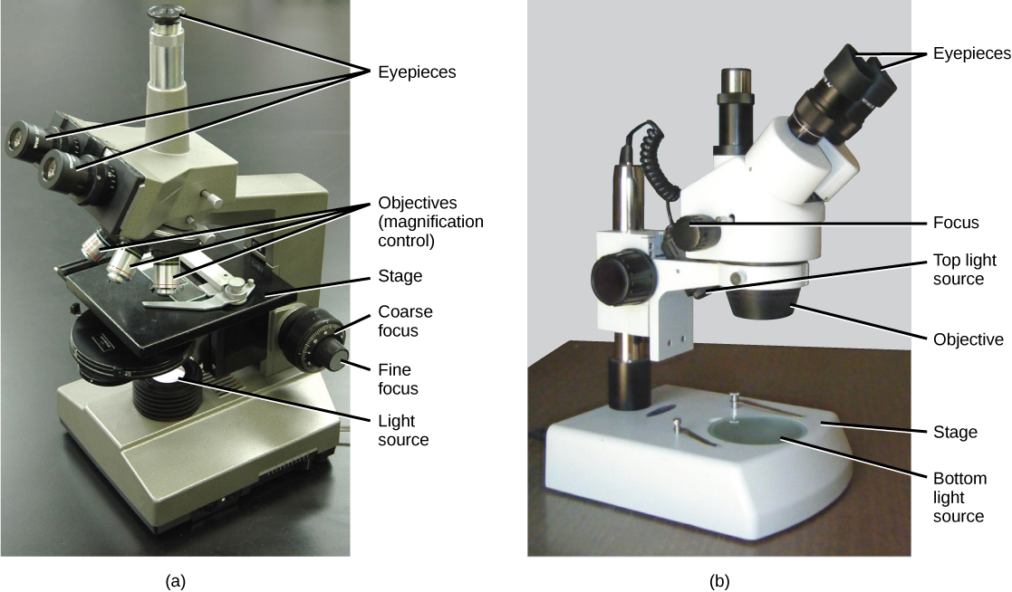
Compound Microscope: Definition, Diagram, Parts, Uses, Working ... - BYJUS Parts of Compound Microscope The compound microscope is mainly used for studying the structural details of cell, tissue, or sections of organs. The parts of a compound microscope can be classified into two: Non-optical parts Optical parts Non-optical parts Base The base is also known as the foot which is either U or horseshoe-shaped.
Drag the labels onto the diagram to identify the parts of the compound microscope (1 of 2).
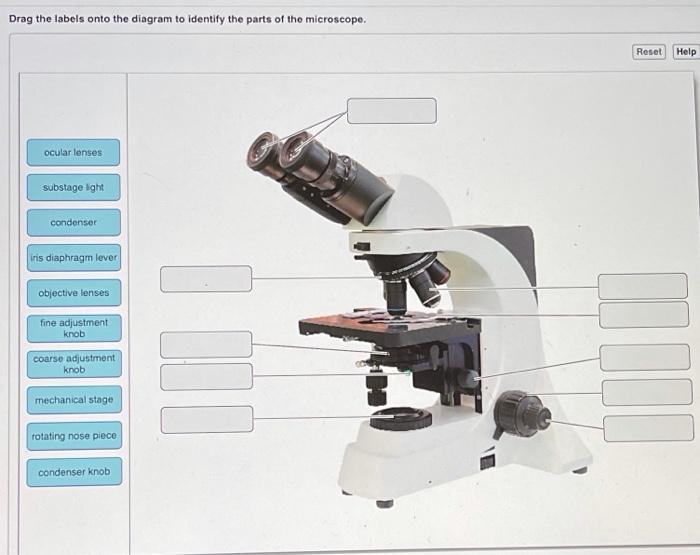
Bio2514 Week 3 The Microscope - Lab Topic.docx - Course Hero Drag the labels onto the diagram to identify the parts of the compound microscope (1 of 2). Arm ocular lens Mechanical stage rotating nose piece Stage Objective lenses Condenser Iris diaphragm lever 3. The microscope slide rests on the __________ while being viewed. Stage 4. Your lab microscope is parfocal. Compound Microscope - Diagram (Parts labelled), Principle and Uses The three structural components include: 1. Head - This is the upper part of the microscope that houses the optical parts 2. Arm - This part connects the head with the base and provides stability to the microscope. Arm is used to carry the microscope around 3. Base - Base is on which the microscope rests and the base houses the illuminator that lights up the specimens Solved Drag the labels onto the diagram to identify the - Chegg Transcribed image text: Drag the labels onto the diagram to identify the parts of the microscope. Reset Help ocular lenses substage ight condenser iris diaphragm lever objective lenses fine adjustment knob coarse adjustment knob mechanical stage rotating nose piece condenser knob Previous question Next question
Drag the labels onto the diagram to identify the parts of the compound microscope (1 of 2).. compound microscope parts (labeling) Flashcards | Quizlet compound microscope parts (labeling) Flashcards | Quizlet compound microscope parts (labeling) Term 1 / 14 eyepiece tube - connects the eyepiece to the objective lens Click the card to flip 👆 Definition 1 / 14 what is 1? Click the card to flip 👆 Flashcards Learn Test Match Created by barnettlily Terms in this set (14) Labelled Diagram of Compound Microscope The below mentioned article provides a labelled diagram of compound microscope. Part # 1. The Stand: The stand is made up of a heavy foot which carries a curved inclinable limb or arm bearing the body tube. The foot is generally horse shoe-shaped structure (Fig. 2) which rests on table top or any other surface on which the microscope in kept. Label the microscope — Science Learning Hub Use this with the Microscope parts activity to help students identify and label the main parts of a microscope and then describe their functions. Drag and drop the text labels onto the microscope diagram. If you want to redo an answer, click on the box and the answer will go back to the top so you can move it to another box. Compound Microscope Parts, Functions, and Labeled Diagram Compound Microscope Definitions for Labels Eyepiece (ocular lens) with or without Pointer: The part that is looked through at the top of the compound microscope. Eyepieces typically have a magnification between 5x & 30x. Monocular or Binocular Head: Structural support that holds & connects the eyepieces to the objective lenses.
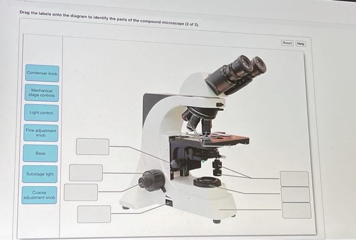
a&p lab 3 hw Flashcards | Quizlet Drag the labels to identify the parts of the compound microscope. Not all labels will be used. 1 left column: head mechanical stage coarse adjustment knob right column: nosepiece stage condenser mechanical stage controls light control Recall from the video the parts of a typical compound microscope. Microscope Parts and Functions Here are the important compound microscope parts... Eyepiece: The lens the viewer looks through to see the specimen. The eyepiece usually contains a 10X or 15X power lens. Diopter Adjustment: Useful as a means to change focus on one eyepiece so as to correct for any difference in vision between your two eyes. Compound Microscope Parts - Labeled Diagram and their Functions There are three major structural parts of a compound microscope. The head includes the upper part of the microscope, which houses the most critical optical components, and the eyepiece tube of the microscope. The base acts as the foundation of microscopes and houses the illuminator. The arm connects between the base and the head parts. Solved Drag the labels onto the diagram to identify the - Chegg Question: Drag the labels onto the diagram to identify the parts of the compound microscope (2 of 2) Malo Condenser knob Mechanical stage controls Light control Fine adjustment knob Base Substage light Coarso adjustmont knob This problem has been solved! You'll get a detailed solution from a subject matter expert that helps you learn core concepts.
16 Parts of a Compound Microscope: Diagrams and Video The 16 core parts of a compound microscope are: Head (Body) Arm Base Eyepiece Eyepiece tube Objective lenses Revolving Nosepiece (Turret) Rack stop Coarse adjustment knobs Fine adjustment knobs Stage Stage clips Aperture Illuminator Condenser Diaphragm Video: Parts of a compound Microscope with Diagram Explained Solved Drag the labels onto the diagram to identify the - Chegg Transcribed image text: Drag the labels onto the diagram to identify the parts of the microscope. Reset Help ocular lenses substage ight condenser iris diaphragm lever objective lenses fine adjustment knob coarse adjustment knob mechanical stage rotating nose piece condenser knob Previous question Next question Compound Microscope - Diagram (Parts labelled), Principle and Uses The three structural components include: 1. Head - This is the upper part of the microscope that houses the optical parts 2. Arm - This part connects the head with the base and provides stability to the microscope. Arm is used to carry the microscope around 3. Base - Base is on which the microscope rests and the base houses the illuminator that lights up the specimens Bio2514 Week 3 The Microscope - Lab Topic.docx - Course Hero Drag the labels onto the diagram to identify the parts of the compound microscope (1 of 2). Arm ocular lens Mechanical stage rotating nose piece Stage Objective lenses Condenser Iris diaphragm lever 3. The microscope slide rests on the __________ while being viewed. Stage 4. Your lab microscope is parfocal.
Disturbance Frequency Determines Morphology and Community ...
Chapter 3: Investigating Proteins – Chemistry
Parts of the Microscope Quiz

Biosensors | November 2022 - Browse Articles

How to Use a Light Microscope: 10 Steps (with Pictures) - wikiHow
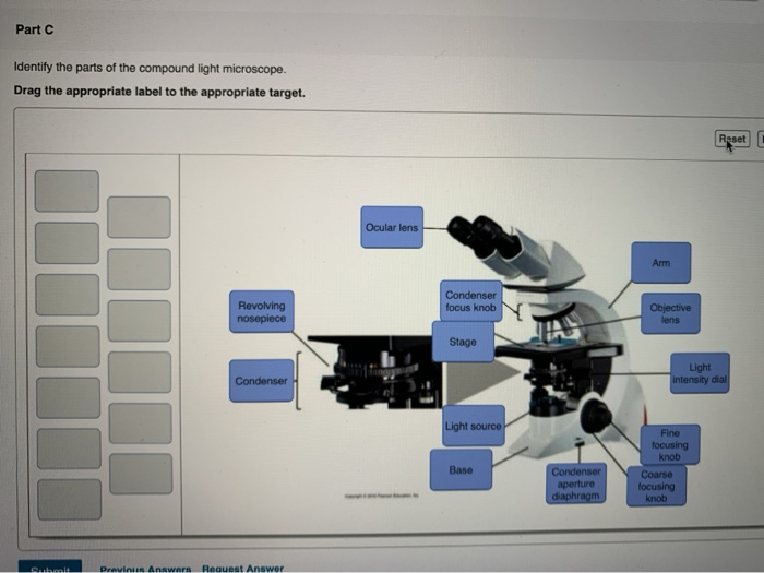
Solved Part C Identify the parts of the compound light ...

Bio2514 Week 3 The Microscope - Lab Topic.docx - Bio2514 Week ...

Electron Microscopy - Laboratorium

Label Microscope Diagram - EnchantedLearning.com
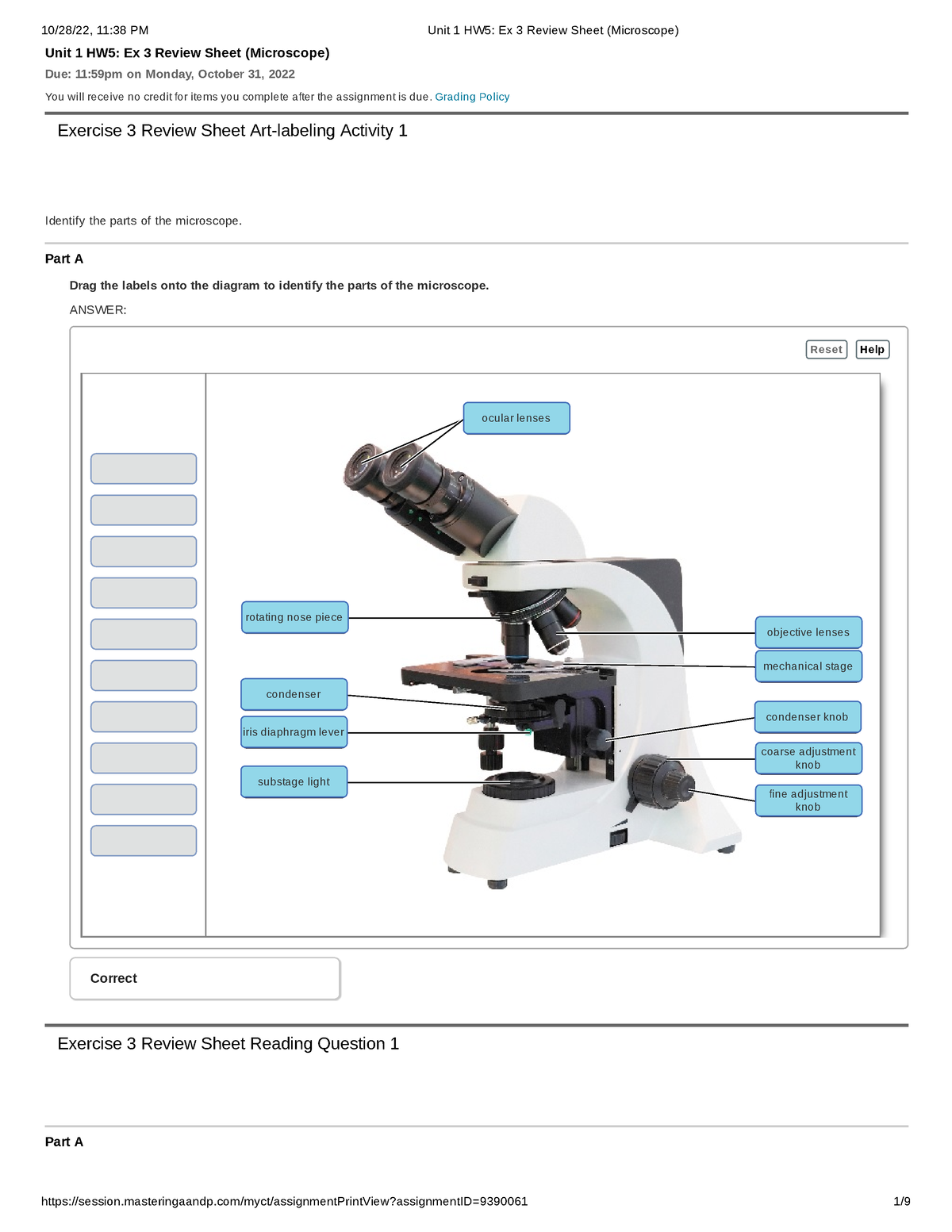
Unit 1 HW5 Ex 3 Review Sheet (Microscope) - Unit 1 HW5: Ex 3 ...
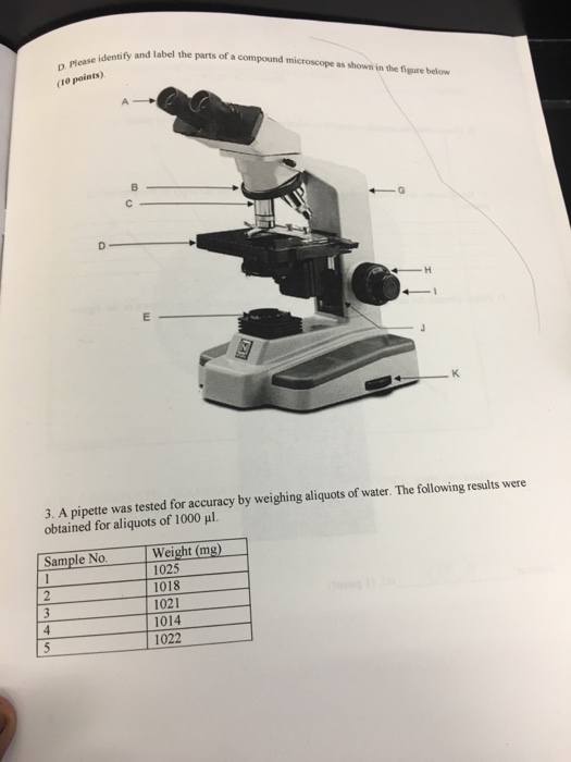
Solved Identify and label the parts of a compound microscope ...

Drag the labels onto the diagram to identify the gustatory ...
Solved Drag the labels onto the diagram to identify the ...

Structure and Function of Blood Vessels - Human Anatomy ...

Microscope Diagram Labeled, Unlabeled and Blank | Parts of a ...

Human Biology

2020 MassURC Research Abstracts | Commonwealth Honors College

Blog | Katie Treggiden

Compound Microscope Parts – Labeled Diagram and their ...

Illustration of the fluorescence spectrum (a) and integrated ...

LAB FINAL Review Flashcards | Quizlet
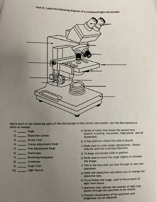
Solved Part III. Label the following diagram of a compound ...

Lab Manual Exercise # 1
BI 122: Biology Lab series (Microscopy module)
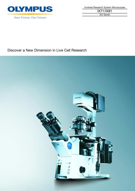
Olympus IX81 Brochure - Microscopes
Label-free imaging and classification of live P. falciparum ...

Label the microscope — Science Learning Hub

Understanding and Developing Science Teachers' PDF | PDF ...

Browse questions for Biology

PAPERmaking! Vol.5 No.1 2019 by pita.co.uk - Issuu

Bio2514 Week 3 The Microscope - Lab Topic.docx - Bio2514 Week ...

Polyelectrolytes in thin liquid films - ScienceDirect
AmScope MU USB3.0 User manual | Manualzz
3.1 How Cells Are Studied – Concepts of Biology – 1st ...

Programmable light-driven swimming actuators via wavelength ...
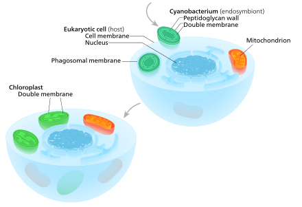
Chloroplast - Wikipedia

Microfluidic Paper-Based Analytical Devices: From Design to ...

Student projects ‒ LIONS ‐ EPFL
LAB 1: Scientific Method/Tools of Scientific Inquiry
Solved Drag the labels onto the diagram to identify the ...
Anatomy of a Microscope | Microscopy Primer | Olympus LS
Post a Comment for "41 drag the labels onto the diagram to identify the parts of the compound microscope (1 of 2)."