43 microscope labeled diagram
Binocular Microscope Anatomy - Parts and Functions with a Labeled Diagram Now, I will discuss the details anatomy of the light compound microscope with the labeled diagram. Why it is called binocular: because it has two ocular lenses or an eyepiece on the head that attaches to the objective lens, this ocular lens magnifies the image produced by the objective lens. Binocular microscope parts and functions label microscope diagram | Charts - Pinterest Feb 26, 2020 - Microscope Diagram - Microscope - Microscope Parts - Diagram of a ... label microscope diagram | Charts Optical Microscope, Microscope Parts, ...
Labeling the Parts of the Microscope - Pinterest Oct 8, 2015 - Microscope World explains the parts of the microscope, including a printable worksheet for schools and home.

Microscope labeled diagram
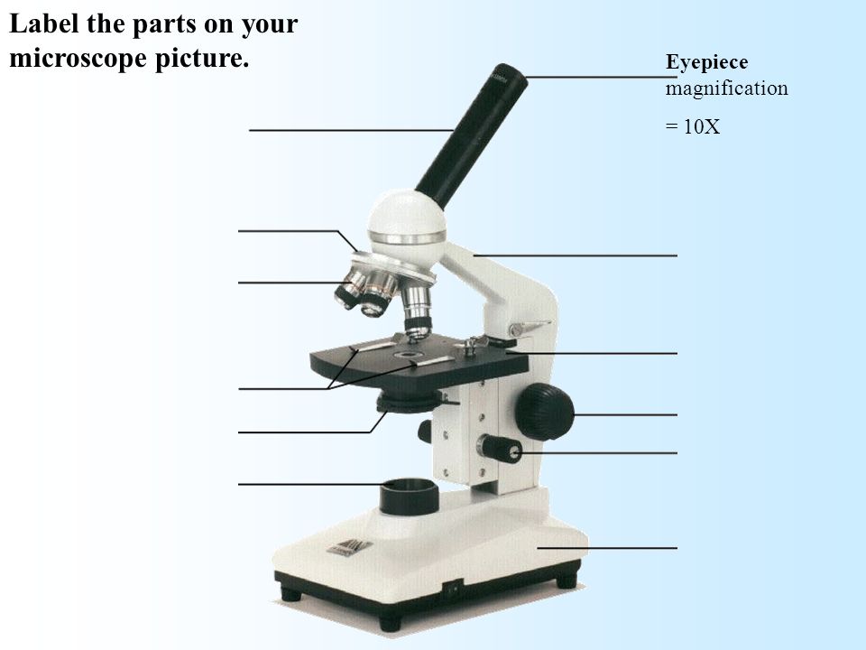
PDF Parts of a Microscope Printables - Homeschool Creations Label the parts of the microscope. You can use the word bank below to fill in the blanks or cut and paste the words at the bottom. Microscope Created by Jolanthe @ HomeschoolCreations.net. Parts of a eyepiece arm stageclips nosepiece focusing knobs illuminator stage objective lenses Compound Microscope Parts, Functions, and Labeled ... Compound Microscope Parts, Functions, and Labeled Diagram Parts of a Compound Microscope Each part of the compound microscope serves its own unique function, with each being important to the function of the scope as a whole. A Study of the Microscope and its Functions ... - Pinterest To better understand the structure and function of a microscope, we need to take a look at the labeled microscope diagrams of the compound and electron ...
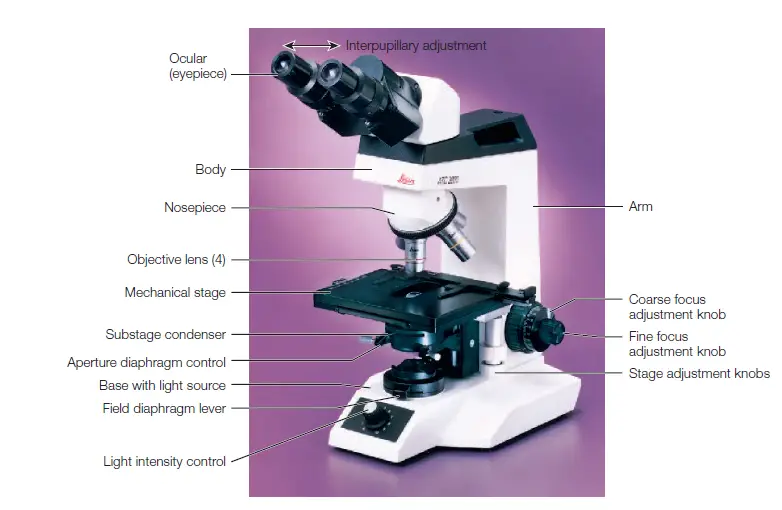
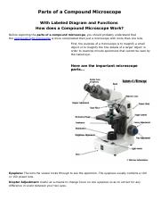
Microscope labeled diagram. Compound Microscope Parts - Labeled Diagram and their Functions Labeled diagram of a compound microscope Major structural parts of a compound microscope There are three major structural parts of a compound microscope. The head includes the upper part of the microscope, which houses the most critical optical components, and the eyepiece tube of the microscope. Compound Microscope - Diagram (Parts labelled), Principle and Uses See: Labeled Diagram showing differences between compound and simple microscope parts Structural Components The three structural components include 1. Head This is the upper part of the microscope that houses the optical parts 2. Arm This part connects the head with the base and provides stability to the microscope. Parts of a microscope with functions and labeled diagram Sep 17, 2022 · Q. Differentiate between a condenser and an Abbe condenser. Ans. Condensers are lenses that are used to collect and focus light from the illuminator into the specimen. They are found under the stage next to the diaphragm of the microscope. They play a major role in ensuring clear sharp images are produced with a high magnification of 400X and above. Microscope: Parts Of A Microscope With Functions And Labeled Diagram. Figure: A diagram of a microscope's components. The microscope has three basic components: the head, the base, and the arm. Head:Occasionally, the head is considered the body. It holds the optical components of the upper part of the microscope. Base:The microscope's base provides great support.
Labeling the Parts of the Microscope Microscope World explains the parts of the microscope, including a printable worksheet for schools and home. Compound Microscope – Diagram (Parts labelled), Principle and … Oct 10, 2022 · See: Labeled Diagram showing differences between compound and simple microscope parts Structural Components. The three structural components include. 1. Head. This is the upper part of the microscope that houses the optical parts. 2. Arm . This part connects the head with the base and provides stability to the microscope. Light Microscope- Definition, Principle, Types, Parts, Labeled Diagram ... Amazing 27 Things Under The Microscope With Diagrams History of Microbiology and Contributors in Microbiology 22 Types of Spectroscopy with Definition, Principle, Steps, Uses Animal Cell- Definition, Structure, Parts, Functions, Labeled Diagram Dark-Field Light Microscope Microscope Parts and Functions With Labeled Diagram and Functions How ... Most specimens are mounted on slides, flat rectangles of thin glass. The specimen is placed on the glass and a cover slip is placed over the specimen. This allows the slide to be easily inserted or removed from the microscope. It also allows the specimen to be labeled, transported, and stored without damage.
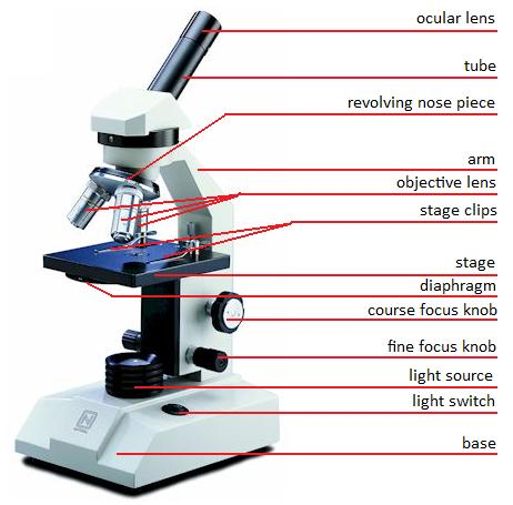
Microscope Types (with labeled diagrams) and Functions Simple microscope labeled diagram Simple microscope functions It is used in industrial applications like: Watchmakers to assemble watches Cloth industry to count the number of threads or fibers in a cloth Jewelers to examine the finer parts of jewelry Miniature artists to examine and build their work Also used to inspect finer details on products Simple Microscope - Diagram (Parts labelled), Principle, Formula and Uses Parts of a Simple Microscope A simple microscope consists of Optical parts Mechanical parts Labeled Diagram of simple microscope parts Optical parts The optical parts of a simple microscope include Lens Mirror Eyepiece Lens A simple microscope uses biconvex lens to magnify the image of a specimen under focus. Parts of a microscope with functions and labeled diagram Figure: Diagram of parts of a microscope There are three structural parts of the microscope i.e. head, base, and arm. Head - This is also known as the body. It carries the optical parts in the upper part of the microscope. Base - It acts as microscopes support. It also carries microscopic illuminators. Labeling the Parts of the Microscope - Pinterest Jan 13, 2016 - Free worksheets for labeling parts of the microscope including a worksheet that is blank and one with answers.
what is that part of microscope. - Brainly.ph
Parts of a microscope with functions and labeled diagram Parts of a microscope with functions and labeled diagram Optical Microscope, Microscope Parts, Digital. microbenotes. Microbe Notes. 2k followers.

Compound and Stereo- microscopes - Microscopes 4 Schools
Microscope, Microscope Parts, Labeled Diagram, and Functions Microscope, Microscope Parts, Labeled Diagram, and Functions What is Microscope? A microscope is a laboratory instrument used to examine objects that are too small to be seen by the naked eye. It is derived from Ancient Greek words and composed of mikrós, "small" and skopeîn,"to look" or "see".

Labelling a Microscope Diagram | Quizlet
Compound Microscope- Definition, Labeled Diagram, Principle, … Apr 03, 2022 · Light Microscope- Definition, Principle, Types, Parts, Labeled Diagram, Magnification Amazing 27 Things Under The Microscope With Diagrams Plant Cell- Definition, Structure, Parts, Functions, Labeled Diagram
Vektor Stok Microscope Diagram Vector Illustration Labeled ...
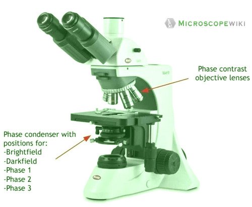
Light Microscope- Definition, Principle, Types, Parts, Labeled Diagram ... Sep 07, 2022 · Figure: Labeled Diagram of a Light Microscope. Types of light microscopes (optical microscope) With the evolved field of Microbiology, the microscopes. used to view specimens are both simple and compound light microscopes, all using lenses. The difference is simple light microscopes use a single lens for magnification while compound lenses use ...
Junior cert Labelling Microscope - Labelled diagram
Microscope With Labels clip art - Pinterest Microscope Diagram Labeled, Unlabeled and Blank | Parts of a Microscope – Tim's Printables. Print a microscope diagram, microscope worksheet, or practice ...
Compound Microscope Parts, Functions, and Labeled Diagram ...
Compound Microscope Parts – Labeled Diagram and their … Major structural parts of a compound microscope. There are three major structural parts of a compound microscope. The head includes the upper part of the microscope, which houses the most critical optical components, and the eyepiece tube of the microscope.; The base acts as the foundation of microscopes and houses the illuminator.; The arm connects between the base …

A labeled diagram of a microscope. MLT 101. :) | Teaching ...
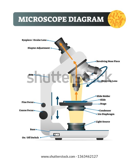
Microscope Parts and Functions Eyepiece: The lens the viewer looks through to see the specimen. The eyepiece usually contains a 10X or 15X power lens. Diopter Adjustment: Useful as a means to change focus on one eyepiece so as to correct for any difference in vision between your two eyes. Body tube (Head): The body tube connects the eyepiece to the objective lenses. Arm: The arm connects the body …
Microscope Types (with labeled diagrams) and Functions
Inverted Microscope- Definition, Principle, Parts, Labeled Diagram ... Apr 10, 2022 · What is an Inverted Microscope? Invented in 1850 by a faculty member of Medical College of Louisiana, named J. Lawrence Smith, this microscope just like it sounds is a light microscope that has its components placed in an inverted order, this means, light source and condenser lens are placed above the specimen stage, pointing down, while the objectives and …
Labeled Parts Of A Microscope - ClipArt Best
parts of a microscope diagram A Study Of The Microscope And Its Functions With A Labeled Diagram . electron anatomy getdrawings. Microscope Labeled Diagram . microscope diagram labeled parts worksheet basic biology science rules using worksheets projects microbiology middle microscopes compound simple blank slideshare printable
Types of Microscopes and Their Uses – Microbe Online
Scanning Electron Microscope (SEM)- Definition, Principle, … Mar 11, 2022 · The first Scanning Electron Microscope was initially made by Mafred von Ardenne in 1937 with an aim to surpass the transmission electron Microscope. He used high-resolution power to scan a small raster using a beam of electrons that were focused on the raster. He also aimed at reducing the problems of chromatic aberrations images produced by the …
Lab :1
Microscope Parts, Function, & Labeled Diagram - slidingmotion Microscope parts labeled diagram gives us all the information about its parts and their position in the microscope. Microscope Parts Labeled Diagram The principle of the Microscope gives you an exact reason to use it. It works on the 3 principles. Magnification Resolving Power Numerical Aperture. Parts of Microscope Head Base Arm Eyepiece Lens

Labeling the Parts of the Microscope | Microscope activity ...
A Study of the Microscope and its Functions With a Labeled Diagram ... A Study of the Microscope and its Functions With a Labeled Diagram To better understand the structure and function of a microscope, we need to take a look at the labeled microscope diagrams of the compound and electron microscope. These diagrams clearly explain the functioning of the microscopes along with their respective parts.

Compound Microscope Parts – Labeled Diagram and their ...
Microscope labeled diagram - SlideShare Microscope labeled diagram Oct. 30, 2013 • 6 likes • 28,032 views Download Now Download to read offline Pisgah High School Follow 1. The Microscope Image courtesy of: Microscopehelp.com Basic rules to using the microscope 1. You should always carry a microscope with two hands, one on the arm and the other under the base. 2.

Microscope Parts and Functions
Microscope label Diagram | Quizlet Start studying Microscope label. Learn vocabulary, terms, and more with flashcards, games, and other study tools.
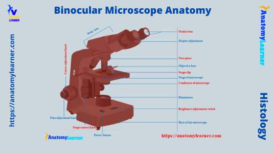
Binocular Microscope Anatomy - Parts and Functions with a ...
Parts of Stereo Microscope (Dissecting microscope) - labeled diagram ... Labeled part diagram of a stereo microscope Major structural parts of a stereo microscope There are three major structural parts of a stereo microscope. The viewing Head includes the upper part of the microscope, which houses the most critical optical components, including the eyepiece, objective lens, and light source of the microscope.

Parts of a Microscope - HaleyMullmicroscopy
Labelled Diagram of Compound Microscope The below mentioned article provides a labelled diagram of compound microscope. Part # 1. The Stand: The stand is made up of a heavy foot which carries a curved inclinable limb or arm bearing the body tube. The foot is generally horse shoe-shaped structure (Fig. 2) which rests on table top or any other surface on which the microscope in kept.

Lable the microscope worksheet
Electron Microscope Principle, Uses, Types and Images (Labeled Diagram ... Ans: A light microscope has a low resolving power (0.25µm to 0.3µm) while the electron microscope has a resolution power about 250 times higher than the light microscope at about 0.001µm. Similarly, a light microscope has a magnification of 500X to 1500x while the electron microscope has a much higher magnification of 100,000X to 300,000X.

Microscope - Mind the Graph
Microscope Parts, Function, & Labeled Diagram - slidingmotion Microscope parts labeled diagram gives us all the information about its parts and their position in the microscope. Microscope Parts Labeled Diagram. The principle of the Microscope gives you an exact reason to use it. It works on the 3 principles. Magnification; Resolving Power; Numerical Aperture. Parts of Microscope.
Parts of a microscope with functions and labeled diagram ...
Label the microscope — Science Learning Hub All microscopes share features in common. In this interactive, you can label the different parts of a microscope. Use this with the Microscope parts activity to help students identify and label the main parts of a microscope and then describe their functions. Drag and drop the text labels onto the microscope diagram.

Compound Microscope Parts, Diagram Definition, Application ...
Microscope, Microscope Parts, Labeled Diagram, and Functions Sep 03, 2022 · Revolving Nosepiece or Turret: Turret is the part of the microscope that holds two or multiple objective lenses and helps to rotate objective lenses and also helps to easily change power. Objective Lenses: Three are 3 or 4 objective lenses on a microscope. The objective lenses almost always consist of 4x, 10x, 40x and 100x powers. The most common eyepiece …

Compound Microscope Parts – Labeled Diagram and their ...
A Study of the Microscope and its Functions ... - Pinterest To better understand the structure and function of a microscope, we need to take a look at the labeled microscope diagrams of the compound and electron ...

Compound Microscope Parts, Functions, and Labeled Diagram ...
Compound Microscope Parts, Functions, and Labeled ... Compound Microscope Parts, Functions, and Labeled Diagram Parts of a Compound Microscope Each part of the compound microscope serves its own unique function, with each being important to the function of the scope as a whole.
Label a microscope - Teaching resources
PDF Parts of a Microscope Printables - Homeschool Creations Label the parts of the microscope. You can use the word bank below to fill in the blanks or cut and paste the words at the bottom. Microscope Created by Jolanthe @ HomeschoolCreations.net. Parts of a eyepiece arm stageclips nosepiece focusing knobs illuminator stage objective lenses

Compound Microscope Parts, Functions, and Labeled Diagram ...

Microscope Labeling Activity - SMART Board Activity - Interactive Review

Simple Microscope Definition, Magnification, Parts And Uses
Microscopes Microscope Parts Quiz on Friday!! - ppt video ...
MICROBIO 16 Parts of a Compound Microscope with Diagram and ...

Microscope With Labels Clip Art at Clker.com - vector clip ...

Parts of the Microscope Label and Definition Diagram | Quizlet

Microscope study part-2

Infographic: Get to Know Your Microscope | Carolina.com

A Study of the Microscope and its Functions With a Labeled ...

Medical Laboratory Science Videos - 💥Brightfield Microscope ...

Draw a labelled diagram of a compound microscope.

Microscope Parts, Types & Diagram | What is a Microscope ...

The Microscope
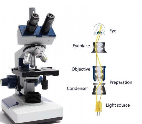
How do microscopes operate? - Krüss laboratory equipment

Compound microscope - their parts and function - Microscopy4kids

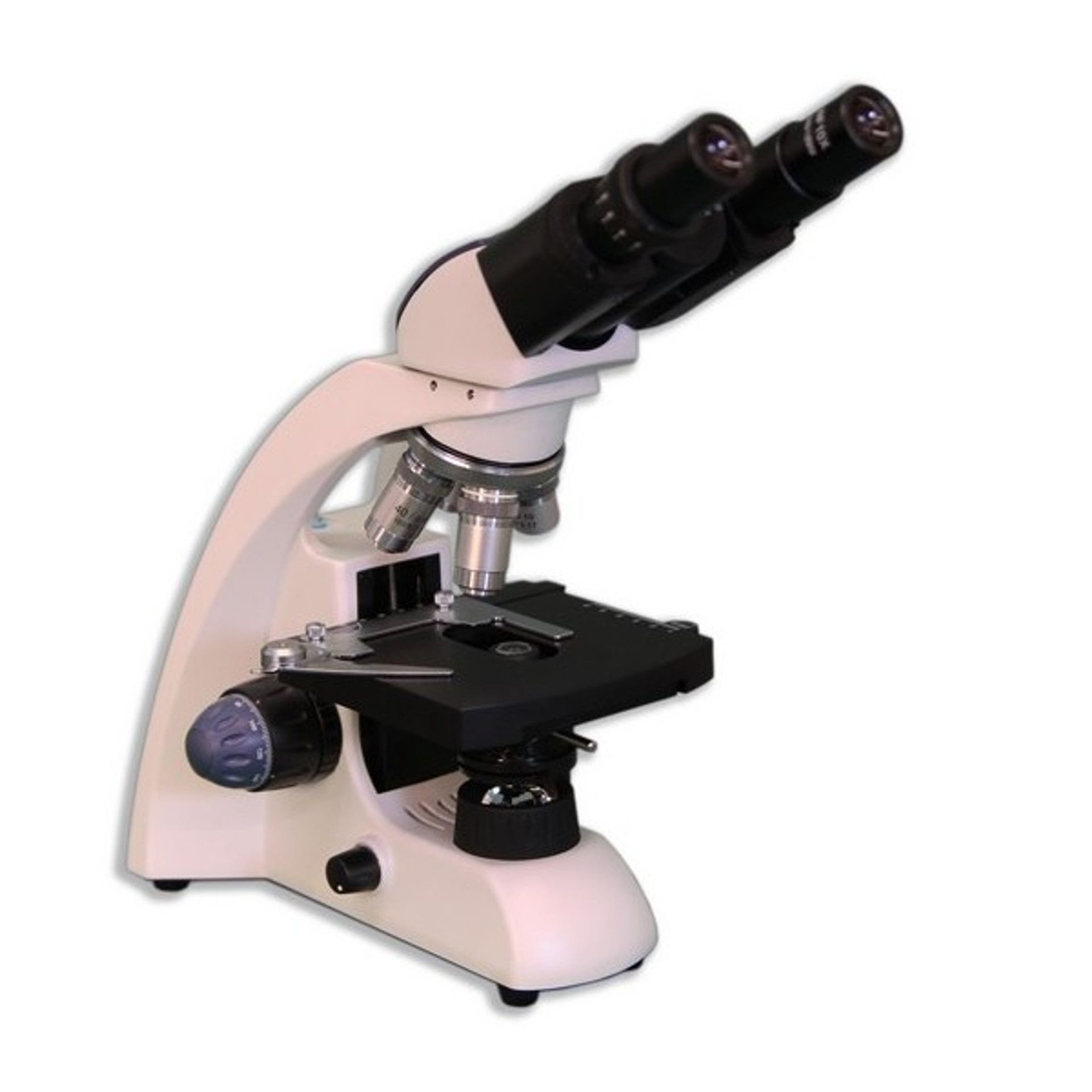
Swift M3600 Series Compound Microscopes: Monocular ...

Draw a neat labelled diagram of a compound microscope class ...
1.2: Microscopes - Biology LibreTexts

Label The Microscope Parts! Diagram | Quizlet
Post a Comment for "43 microscope labeled diagram"