42 diagram of a microscope with label
Compound Microscope - Types, Parts, Diagram, Functions and Uses It comes with a wide body and base. Its distinct parts include a condenser, illumination, focus lock, mechanical stage, and a revolving nosepiece which can hold up to five objectives. It usually has a binocular head, which makes long-term observation easy. Image 22: An example of a research compound microscope. Microscope: Types of Microscope, Parts, Uses, Diagram - Embibe There microscope anatomy includes three structural parts, i.e. head, base, and arm. Head - This is also known as the body; it carries the optical parts in the upper part of the microscope.. Base - It acts as microscopes support.It also carries microscopic illuminators. Arms - The microscope arm connects the base and the head and the eyepiece tube to the microscope base.
Microscopy- History, Classification, Terms, Diagram - The Biology Notes History of Microscope. In the 1 st Century AD, the Romans invented the glass and used them to magnify objects. In the early 14 th Century AD, eyeglasses were made by Italian spectacle makers. In 1590, two Dutch spectacle makers, Hans, and Zacharias Jansen created the first microscope. It was a simple tube with 2 lenses system and had 9X ...

Diagram of a microscope with label
Microscope, Microscope Parts, Labeled Diagram, and Functions The description given below summarize the brief description of microscope parts used to visualize the microscopic specimens such as animal cells, plant cells, microbes, bacteria, viruses, microorganisms etc. The Microscopes parts divided into three different structural parts Head, Base, and Arms. Microscope Diagram Worksheet - The Microscope Create A Labelled Diagram ... Microscope Labeled Diagram from cdn.slidesharecdn.com Used to support the microscope when carried. There is a printable worksheet available for download here so you can take the . This online quiz is called microscope labeling game science, microsope. Be sure to check our teachers notebook store for other printables. Microscope Parts, Function, & Labeled Diagram - slidingmotion Microscope parts labeled diagram gives us all the information about its parts and their position in the microscope. Microscope Parts Labeled Diagram The principle of the Microscope gives you an exact reason to use it. It works on the 3 principles. Magnification Resolving Power Numerical Aperture. Parts of Microscope Head Base Arm Eyepiece Lens
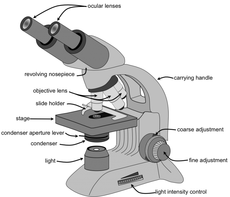
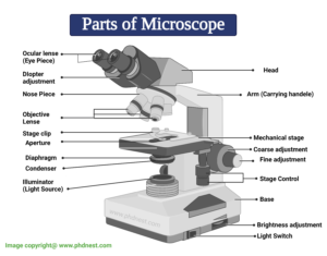
Diagram of a microscope with label. Parts of a microscope with functions and labeled diagram - Microbe Notes Q. List down the 18 parts of a Microscope. 1. Ocular Lens (Eye Piece) 2. Diopter Adjustment 3. Head 4. Nose Piece 5. Objective Lens 6. Arm (Carrying Handle) 7. Mechanical Stage 8. Stage Clip 9. Aperture 10. Diaphragm 11. Condenser 12. Coarse Adjustment 13. Fine Adjustment 14. Illuminator (Light Source) 15. Stage Controls 16. Base 17. Blood Histology Slides with Description and Labeled Diagram The blood is a specialized connective tissue that is fluid and circulates through the vascular channel. In the blood histology slide, you will find different types of cells with their specific features. This might be a short article where I will show you all the cells from the blood microscope slide with a labeled diagram and actual pictures. Inverted Microscope- Definition, Principle, Parts, Labeled Diagram ... Uses of the Inverted Microscope. It is used in diagnostics fungal cultures, for example, detection of Phytophthora spp in cultures. Used for diagnosis of nematology extraction specimens to observe nematodes such as Vermiform nematodes. Used to observe living microbial cells found at the bottom of lab vessels such as tissue culture flasks and ... Binocular Microscope Anatomy - Parts and Functions with a Labeled Diagram The nose piece of a microscope, Head part of the microscope, Ocular lens or eyepiece of the microscope, Diopter adjustment of the eyepiece All of these parts are identified in a light microscope labeled diagram. So, first, make sure you can identify all these parts from this labeled diagram. Parts of the compound microscope
Draw a diagram of a matured microspore of an angiosperm. Label its ... Draw a diagram of L.S. of an anatropous ovule of an angiosperm and label the following parts. asked Aug 17, 2021 in Biology by Devakumari ( 52.3k points) sexual reproduction in flowering plants Microscope Types (with labeled diagrams) and Functions Phase-contrast microscope labeled diagram Phase-contrast microscope functions: Its applications areas include In cases where the specimen is colorless and is very tiny In biology to conduct cellular level examination of microorganisms that can't be visualized using the bright field microscopy Interference Microscope Sperm Under Microscope with Labeled Diagram - AnatomyLearner Sperm Under Microscope with Labeled Diagram 24/06/2022 17/06/2022 by anatomylearner While studying the histological features of the seminiferous tubules and epididymis, you will see sperm cells under the microscope. They are much smaller and lie in groups along the inner margin of the Sertoli cells. Simple Microscope - Parts, Functions, Diagram and Labelling Stage - The stage of the microscope is a metal plate that is rectangular in shape and fitted to the vertical rod. It comes with a hole in the center that enables the light to pass from below. The stage holds the slide that contains the specimen to be examined for.
Compound Microscope - Diagram (Parts labelled), Principle and Uses See: Labeled Diagram showing differences between compound and simple microscope parts Structural Components The three structural components include 1. Head This is the upper part of the microscope that houses the optical parts 2. Arm This part connects the head with the base and provides stability to the microscope. Compound Microscope- Definition, Labeled Diagram, Principle, Parts, Uses A standard Microscope has three to four Objective Lenses which range from 4X to 100X. Stage Clips are metal clips that held the slide in place. Arm and Base The Arm connects the Body Tube to the base of the Microscope. The Base supports the Microscope and its where Illuminator. Illuminator and Stage Light Microscope- Definition, Principle, Types, Parts, Labeled Diagram ... Amazing 27 Things Under The Microscope With Diagrams Parts of a microscope with functions and labeled diagram 22 Types of Spectroscopy with Definition, Principle, Steps, Uses History of Microbiology and Contributors in Microbiology Microbiology of extreme environments (Types and Examples) Dark-Field Light Microscope Simple Squamous Epithelium under a Microscope with a Labeled Diagram ... Here the artery labeled diagram shows the tunica intima that consists of endothelium, basal lamina, subendothelium connective tissue, and internal elastic lamina. You will find the endoplasmic reticulum and mitochondria in the cytoplasm of the endothelium cell under the electron microscope.
LABELING THE COMPOUND LIGHT MICROSCOPE 2 Diagram | Quizlet
Simple Microscope - Diagram (Parts labelled), Principle, Formula and Uses Labeled Diagram of simple microscope parts Optical parts. The optical parts of a simple microscope include. Lens; Mirror; Eyepiece; Lens. A simple microscope uses biconvex lens to magnify the image of a specimen under focus.

parts of microscope with diagram
Microscope Diagram - slide preparation biology4isc, anthrax microbewiki ... Microscope Diagram - 15 images - lycopodium, cell division of e coli with continuous media flow youtube, labelled microscope diagram gcse micropedia, give a well labelled diagram of compound microscope using of typical,

Compound Microscope- Definition, Labeled Diagram, Principle ...
Electron Microscope Principle, Uses, Types and Images (Labeled Diagram ... Electron Microscope Principle, Uses, Types and Images (Labeled Diagram), Price Electron Microscope The advances in technology have enabled the development of powerful microscopes to view the samples at a nanometer level and thus were born the electron microscopes.
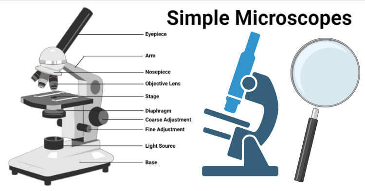
Simple Microscope- Definition, Principle, Magnification ...
Parts of the Microscope with Labeling (also Free Printouts) Parts of the Microscope with Labeling (also Free Printouts) By Editorial Team March 7, 2022 A microscope is one of the invaluable tools in the laboratory setting. It is used to observe things that cannot be seen by the naked eye. Table of Contents 1. Eyepiece 2. Body tube/Head 3. Turret/Nose piece 4. Objective lenses 5. Knobs (fine and coarse) 6.

How to draw compound of Microscope easily - step by step
Electron Microscope-Definition, Principle, Types, Uses, Labeled Diagram The electron microscope is placed vertically and has the shape of a tall vacuum column. It consists of the following elements: 1. Electron gun. A heated tungsten filament that produces electrons makes up the electron cannon. 2. Electromagnetic lenses. The condenser lens directs the electron beam to the specimen.

MICROSCOPE Labeling - Part - 3
Parts of a Microscope: Lesson for Kids - Study.com Instructor: Emily Lockhart. Emily has taught science and has a master's degree in education. Learn about the parts of a microscope to see how they help us see small things. Explore the ocular lens ...

Compound Microscope Parts – Labeled Diagram and their ...
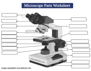
Label The Parts Of A Microscope Worksheet Answers You can use the word bank below to fill in the blanks or cut. Label the parts of a microscope worksheet answers. Students label the parts of the microscope in this photo of a basic laboratory light microscope. Files include a link to editable doc so you can rewrite a. Power 10 x 4 40 Power 10 x 10 100 Power 10 x 40 400 What happens as the power ...
22 Parts Of a Microscope With Their Function And Labeled ...
Neuron under Microscope with Labeled Diagram - AnatomyLearner But, first, let's try to identify the following features from a neuron with the help of a labelled diagram. Cell body or perikaryon of a neuron Nucleus, cytoplasm, the plasma membrane of a neuron Nissl bodies in the cell body of a neuron An initial segment of axon and axon hillock Dendrites and axons of a neuron Axolemma and myelin sheath

Label Microscope Diagram - EnchantedLearning.com
Plant Cell Under Microscope 40X Labeled - Blogger See how a generalized structure of an animal cell and plant cell look with labeled diagrams. Under the microscope, plant cells are seen as large rectangular interlocking blocks. Set up your microscope, place the onion root slide on the stage and focus on low (40x) power. 3) to draw and label a plant cell under 40x, a spider under 4x and human ...
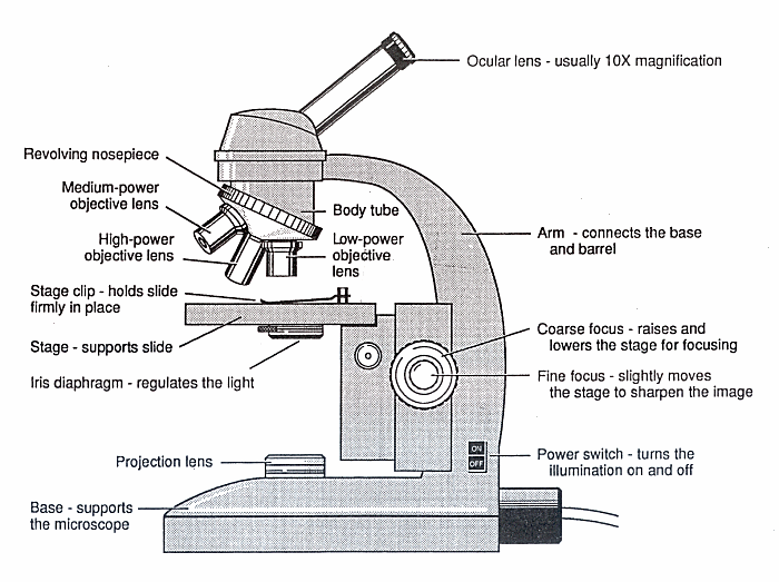
Diagram of a Microscope - Guide to using a microscope
Bright-field microscope (Compound light microscope) - Diagram (Parts ... Bright-field microscope parts (Labeled Diagram) Ocular Lens This microscope has two eye lenses or ocular lens on the top of the microscope that are used to focus the image from the objective lens. It is from these lenses that we see the magnified image of the specimen. Objective Lens
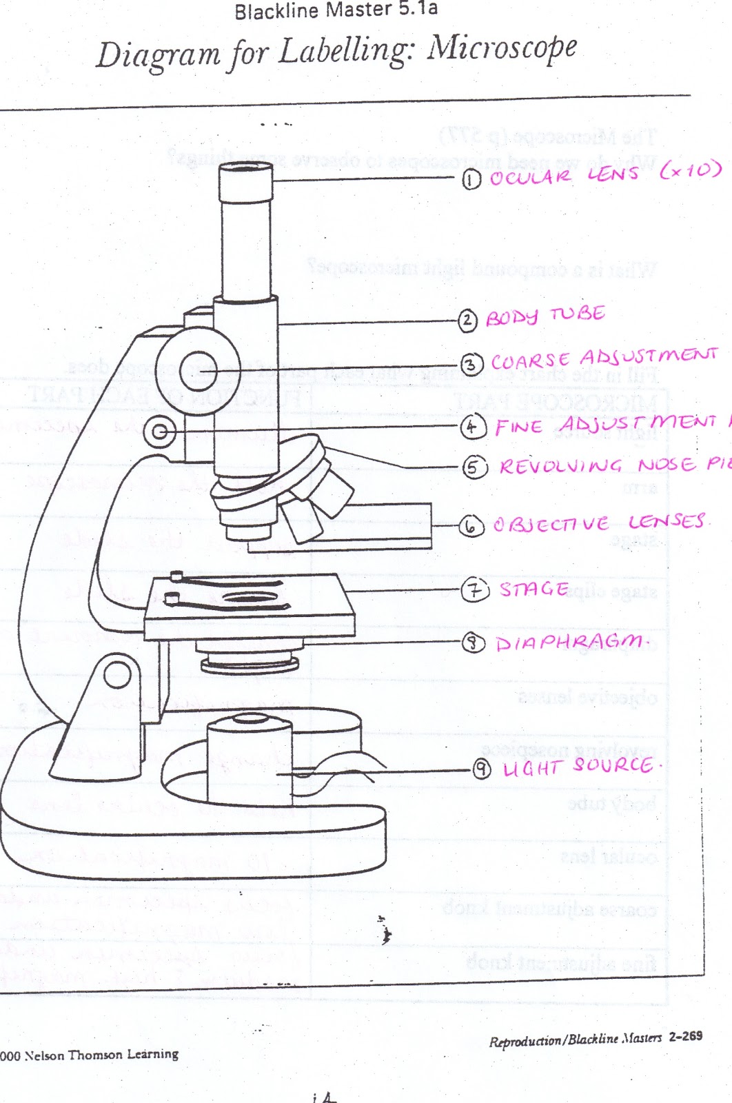
All Saints Online: Diagram for Labelling: Microscope
Electron Microscope- Definition, Principle, Types, Uses, Labeled Diagram Parts of Electron Microscope Electron Microscope is in the form of a tall vacuum column that is vertically mounted. It has the following components: 1. Electron gun The electron gun is a heated tungsten filament, which generates electrons. 2. Electromagnetic lenses The condenser lens focuses the electron beam on the specimen.

Simple Microscope Definition, Magnification, Parts And Uses
Microscope Parts, Function, & Labeled Diagram - slidingmotion Microscope parts labeled diagram gives us all the information about its parts and their position in the microscope. Microscope Parts Labeled Diagram The principle of the Microscope gives you an exact reason to use it. It works on the 3 principles. Magnification Resolving Power Numerical Aperture. Parts of Microscope Head Base Arm Eyepiece Lens

Labeling the Parts of the Microscope | Microscope World Resources
Microscope Diagram Worksheet - The Microscope Create A Labelled Diagram ... Microscope Labeled Diagram from cdn.slidesharecdn.com Used to support the microscope when carried. There is a printable worksheet available for download here so you can take the . This online quiz is called microscope labeling game science, microsope. Be sure to check our teachers notebook store for other printables.

Pin page
Microscope, Microscope Parts, Labeled Diagram, and Functions The description given below summarize the brief description of microscope parts used to visualize the microscopic specimens such as animal cells, plant cells, microbes, bacteria, viruses, microorganisms etc. The Microscopes parts divided into three different structural parts Head, Base, and Arms.

Parts of a microscope with functions and labeled diagram

Lable the microscope worksheet

Compound Microscope: Parts of Compound Microscope
Bright Field Microscope: Definition, Parts, Diagram ...

This is a common compound microscope. Label its parts from A ...
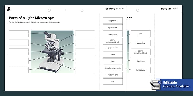
Parts of a Light Microscope Cut and Stick Worksheet - Twinkl
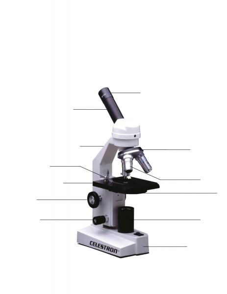
Name Date Sci STANDARD MICROSCOPE DIAGRAM Label only the ...
Labeled Microscope Storyboard by oliversmith
Print Map Quiz: Labeling the Microscope ()

The Microscope
Parts of Microscope, Microscope Labeled Diagram and Functions ...

Microscope Parts and Functions

Microscope Diagram Labeled, Unlabeled and Blank | Parts of a ...

Produk Microscope | UD Berkah Abadi

Compound Microscope Parts

Microscope With Labels Clip Art at Clker.com - vector clip ...

Parts of a microscope with functions and labeled diagram
Labeling Microscope Worksheet | Teaching Resources

Microscope Diagram – Charts

Labelling a Microscope Diagram | Quizlet
Modified Science Diagram; Label Parts of a Microscope; Special Education

microscope parts LABELED

microscope drawing with label - Clip Art Library

Draw a labelled diagram of a compound microscope.

Microscope Diagram - Label Diagram | Quizlet

Simple Microscope - Diagram (Parts labelled), Principle ...

Parts of a Microscope - SmartSchool Systems

Parts of a Microscope with Their Functions • Microbe Online
Post a Comment for "42 diagram of a microscope with label"