39 standard microscope labeled
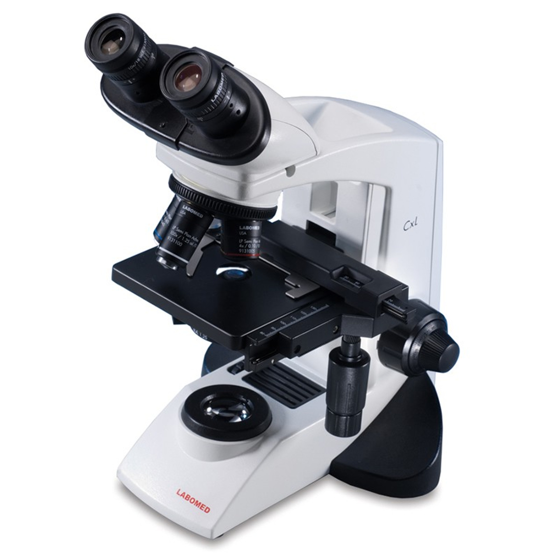
Microscope slide - Wikipedia A set of standard 75 by 25 mm microscope slides. The white area can be written on to label the slide. A microscope slide (top) and a cover slip (bottom) A microscope slide is a thin flat piece of glass, typically 75 by 26 mm (3 by 1 inches) and about 1 mm thick, used to hold objects for examination under a microscope. What do the numbers on the barrel of the microscope ... - Celestron November 24, 2008. Microscope objective lenses will often have four numbers engraved on the barrel in a 2x2 array. The upper left number is the magnification factor of the objective. For example, 4x, 10x, 40x, and 100x. The upper right number is the numerical aperture of the objective. For example 0.1, 0.25, 0.65, and 1.25.
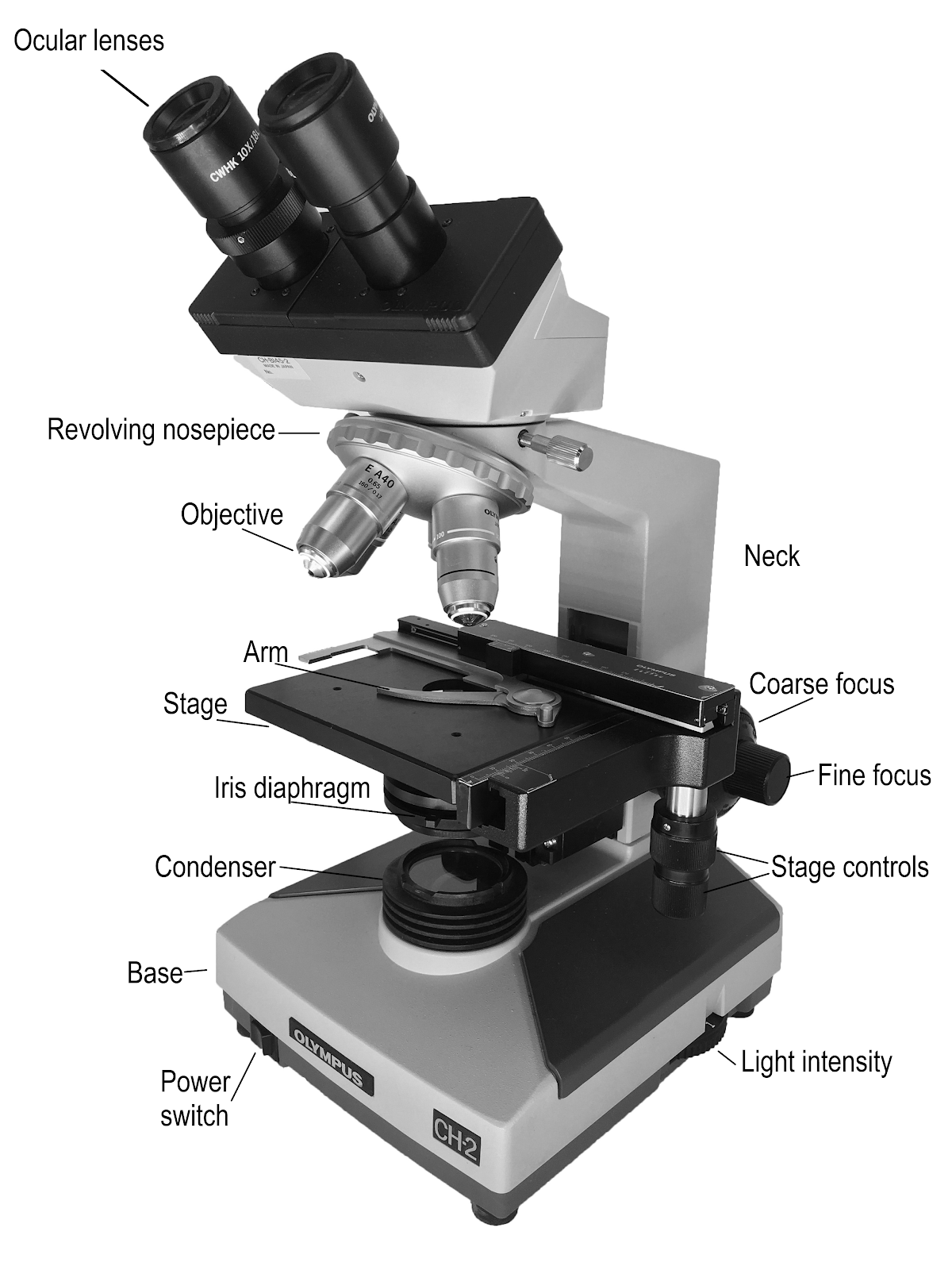
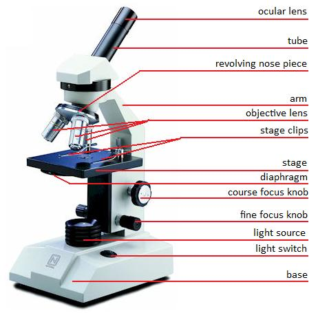
Parts of the Microscope with Labeling (also Free Printouts) Parts of the Microscope with Labeling (also Free Printouts) By Editorial Team March 7, 2022 A microscope is one of the invaluable tools in the laboratory setting. It is used to observe things that cannot be seen by the naked eye. Table of Contents 1. Eyepiece 2. Body tube/Head 3. Turret/Nose piece 4. Objective lenses 5. Knobs (fine and coarse) 6.
Standard microscope labeled
Label the microscope — Science Learning Hub All microscopes share features in common. In this interactive, you can label the different parts of a microscope. Use this with the Microscope parts activity to help students identify and label the main parts of a microscope and then describe their functions. Drag and drop the text labels onto the microscope diagram. Microscope Parts and Functions A standard microscope has three, four, or five objective lenses that range in power from 4X to 100X. When focusing the microscope, be careful that the objective lens doesn't touch the slide, as it could break the slide and destroy the specimen. Specimen or slide: The specimen is the object being examined. Compound Microscope Parts, Functions, and Labeled Diagram Compound Microscope Definitions for Labels. Eyepiece (ocular lens) with or without Pointer: The part that is looked through at the top of the compound microscope. Eyepieces typically have a magnification between 5x & 30x. Monocular or Binocular Head: Structural support that holds & connects the eyepieces to the objective lenses.
Standard microscope labeled. Parts of a microscope with functions and labeled diagram - Microbe Notes Its found at the top of the microscope. Its standard magnification is 10x with an optional eyepiece having magnifications from 5X to 30X. Eyepiece tube - it's the eyepiece holder. It carries the eyepiece just above the objective lens. Stereo Microscope - Parts, Types and Uses - Laboratoryinfo.com Stereo Microscope Parts and Functions. It has three key parts namely: body, focus block, and viewing head/body. Let us take a look at the functions of every part.. Body/viewing head - It houses the optical parts in the upper section of the microscope. Focus block - It attaches the head of the microscope to the stand and focuses it. Stand - It supports the microscope as well as houses ... Understanding Microscopes and Objectives | Edmund Optics Understanding Microscopes and Objectives. A microscope is an optical device used to image an object onto the human eye or a video device. The earliest microscopes, consisting of two elements, simply produced a larger image of an object under inspection than what the human eye could observe. The design has evolved over the microscope's history ... Microscope Slide Labels - Histology Labels - adazonusa.com Custom Microscope Slide Labels. Adazon offers microscope slide labels in a variety of materials, sizes and shapes to meet the needs of your laboratory. Choose blank or pre-printed thermal transfer labels. Our microscope slide labels are available in standard (thin) or pathology thickness. We offer square or rounded corners with a permanent ...
Stereo Microscope Parts Set the microscope on a flat surface in a stable and comfortable position. Turn on The Transmitted/Oblique illuminator. Place a small solid specimen onto the stage such as a card, coin or any other flat, detailed object. Turn the Magnification adjustment knob to the lowest power and bring the image into focus using the focus control. 22 Parts Of a Microscope With Their Function And Labeled Diagram A microscope is a laboratory instrument used to examine objects that are too small to be seen by the naked eye. In other words, it enlarges images of small objects. Invented by a Dutch spectacle maker in the late 16th century, light microscopes use lenses and light to magnify images. Anatomy of the Microscope - Microscope Stages | Olympus LS A simple (commonly termed "plain") microscope stage is illustrated on the left in Figure 2. This stage contains an opening to admit light from the condenser, several mounting holes for a mechanical stage, and two clips that secure the specimen slide in place for observation under increasing magnification (changing of objectives) and for photomicrography. Parts of Stereo Microscope (Dissecting microscope) - labeled diagram ... Labeled part diagram of a stereo microscope Major structural parts of a stereo microscope. ... The eyepiece (or ocular lens) is the lens part at the top of a microscope that the viewer looks through. Typically, standard eyepieces for a dissecting microscope have a magnifying power of 10x. Optional eyepieces of varying powers are available ...
Standard Microscope Slides - daigger.com The Daigger Standard Microscope Slides are made from corrosion-resistant glass with smoothly polished edges that won't snag gloves and measure 75 x 25 mm (3" x 1"), with an approximate thickness of 1 mm. Precleaned; packages 72 slides per box; 20 boxes per case.. Key Feature . Excellent for educational use; Glass is corrosion-resistant and has polished edges Microscope Magnification: Explained - Microscope Clarity The objective lens magnification power is usually displayed prominently as a number and then an "X" or the number before the slash. The objective lenses are also color coded. Red is the lowest power, yellow the next highest power, and blue is the highest power on a microscope with three objectives. Limits to Magnification (Empty Magnification) Labeling of Objectives | Products | Leica Microsystems Each objective is labeled with its magnification, for instance 5x or 100x. However, the magnification of the objective alone does not determine the overall magnification of the microscope. This results from the objective magnification multiplied by the eyepiece magnification (for tube lens 1x). Example: Light Microscope- Definition, Principle, Types, Parts, Labeled Diagram ... Parts of a microscope with functions and labeled diagram 22 Types of Spectroscopy with Definition, Principle, Steps, Uses History of Microbiology and Contributors in Microbiology Microbiology of extreme environments (Types and Examples) Dark-Field Light Microscope
Microscope, Microscope Parts, Labeled Diagram, and Functions
Microscope Slide Labels | Quality Materials | Order at PDC StainerShield® Slide Label Stain Resistant - Direct Thermal Synthetic Permanent 1" Core 7/8" x 7/8" White, 7200 per Roll, 1 Roll per Box. TDSS1-4-7878. $1,146.26. Add to Cart. Compare. StainerShield® Slide Label Stain Resistant - Direct Thermal Synthetic Permanent 3" Core 7/8" x 7/8" White, 1800 per Roll, 4 Rolls per Box. TDSS3-1-7878.

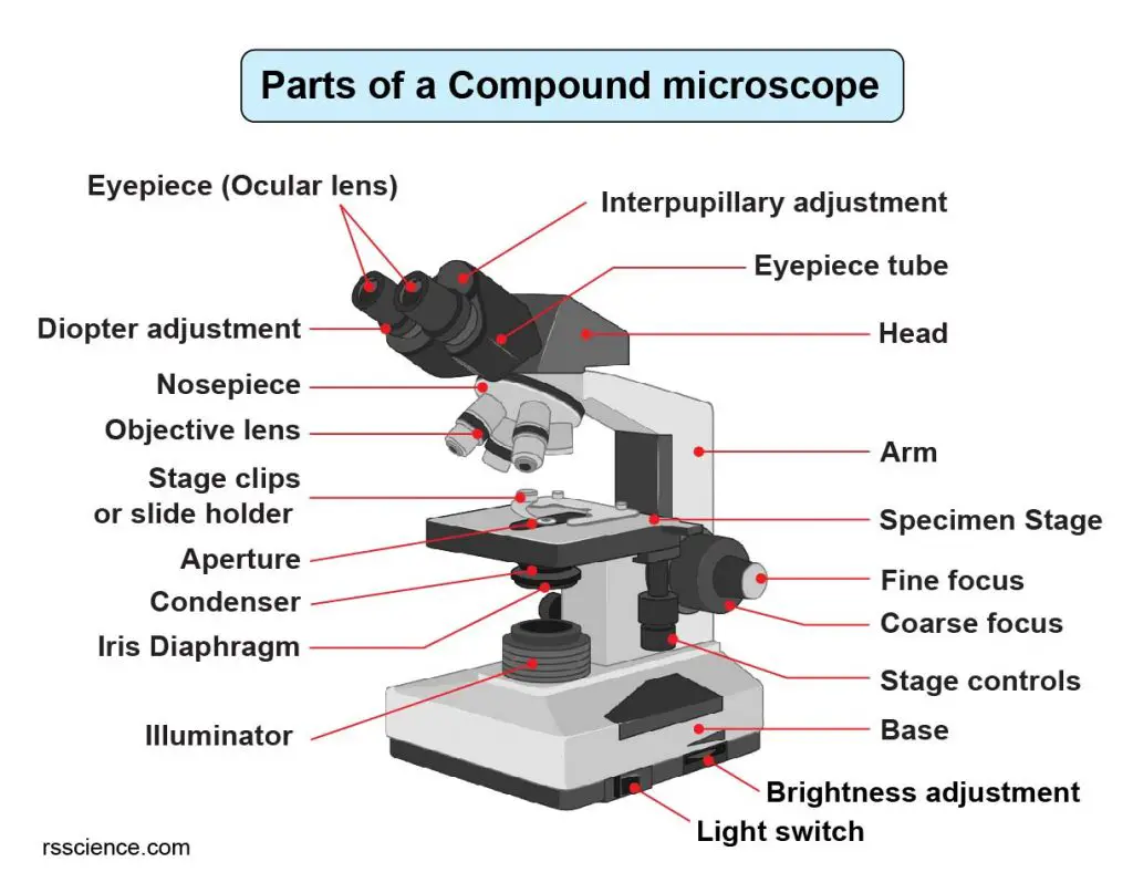
Parts of a microscope with functions and labeled diagram
PDF Label parts of the Microscope: Answers Label parts of the Microscope: Answers Coarse Focus Fine Focus Eyepiece Arm Rack Stop Stage Clip . Created Date: 20150715115425Z ...

Compound Microscope Parts – Labeled Diagram and their ...
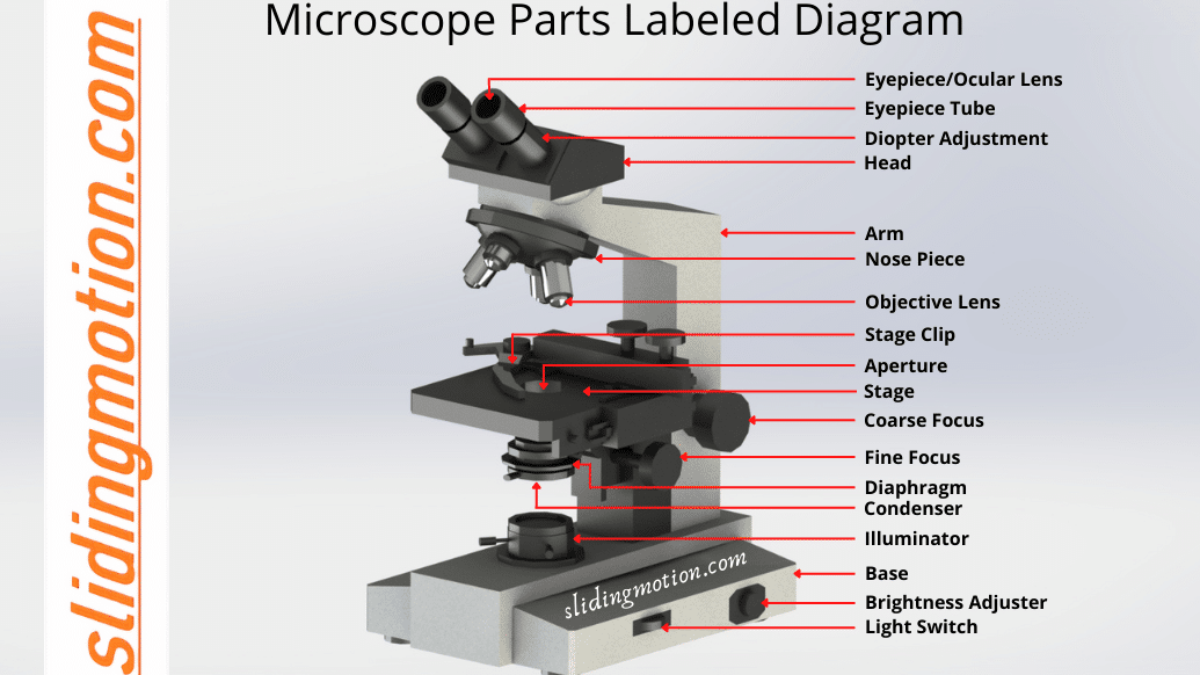
Microscope Parts, Function, & Labeled Diagram - slidingmotion Microscope Parts Labeled Diagram The principle of the Microscope gives you an exact reason to use it. It works on the 3 principles. Magnification Resolving Power Numerical Aperture. Parts of Microscope Head Base Arm Eyepiece Lens Eyepiece Tube Objective Lenses Nose Piece Adjustment Knobs Stage Aperture Microscopic Illuminator Condenser Lens

Amazon.com: SWIFT Microscope SW150,Compound Student ...
Microscope Parts & Functions - AmScope Objective Lenses: Usually you will find 3 or 4 objective lenses on a microscope. The most common ones are 4X (shortest lens), 10X, 40X and 100X (longest lens). The higher power objectives (starting from 40x) are spring loaded.

Microscope With Labels clip art | Microscope parts ...
Compound Microscope Parts - Labeled Diagram and their Functions The eyepiece (or ocular lens) is the lens part at the top of a microscope that the viewer looks through. The standard eyepiece has a magnification of 10x. You may exchange with an optional eyepiece ranging from 5x - 30x. [In this figure] The structure inside an eyepiece. The current design of the eyepiece is no longer a single convex lens.

Microscope Parts and Functions
DIN Standard Microscope Objective Lenses The focal tube length of a DIN standard microscope objective is 160mm. A typical DIN standard microscope objective lens has 0.7965" (20.1mm) diameter threads, 36 TPI (threads per inch), and 55° whitworth. The former standard was RMS ("Royal Microscope Society"), which had a longer tube length (170mm). Most DIN optics are interchangeable.

SWIFT SW380T Siedentopf Trinocular Compound Microscope for Adults,With 40X-2500X And Double-Layer Mechanical Stage
Types of Microscopes: Definition, Working Principle, Diagram ... - BYJUS A compound microscope is defined as the type of microscope that has more than one lens. It has a combination of lenses and two optical parts known as an objective lens and an eyepiece or ocular lens. The magnifying power of the compound microscope is given as: m = D f o × L f e Where, D is the least distance of distinct vision

2.1 " Compound Microscope" | Download Scientific Diagram
A Study of the Microscope and its Functions With a Labeled Diagram ... Objective Lenses - A standard compound microscope contains two primary objective lenses, which can have a magnification of 4x, 5x, 10x, 20x, 40x, 50x, and 100x. The magnification values are written on the side of each lens.

Solved A. OLYMPUS C. B. Use the Diagram to answer the | Chegg.com
Anatomy of the Microscope - Objectives: Specifications and ... - Olympus The thickness of these small glass plates is now standardized at 0.17 mm for most applications, although there is often some variation in thickness within a batch of coverslips.
Microscope labeled diagram
Microscope, Microscope Parts, Labeled Diagram, and Functions There is various type of microscope such as transmission electron microscopes (TEMs), scanning electron microscopes (SEMs), atomic force microscopes (AFM), near-field scanning optical microscopes (MSOM or SNOM, scanning near-field optical microscopy, and scanning tunneling microscopes (STM).

E-Katalog 5.0
PDF Parts of the Light Microscope - Science Spot B. NOSEPIECE microscope when carried Holds the HIGH- and LOW- power objective LENSES; can be rotated to change MAGNIFICATION. Power = 10 x 4 = 40 Power = 10 x 10 = 100 Power = 10 x 40 = 400 What happens as the power of magnification increases?

File:Labelledmicroscope.gif - Wikimedia Commons
Compound Microscope Parts, Functions, and Labeled Diagram Compound Microscope Definitions for Labels. Eyepiece (ocular lens) with or without Pointer: The part that is looked through at the top of the compound microscope. Eyepieces typically have a magnification between 5x & 30x. Monocular or Binocular Head: Structural support that holds & connects the eyepieces to the objective lenses.
What does the objective lens do on a microscope? - Dr ...
Microscope Parts and Functions A standard microscope has three, four, or five objective lenses that range in power from 4X to 100X. When focusing the microscope, be careful that the objective lens doesn't touch the slide, as it could break the slide and destroy the specimen. Specimen or slide: The specimen is the object being examined.
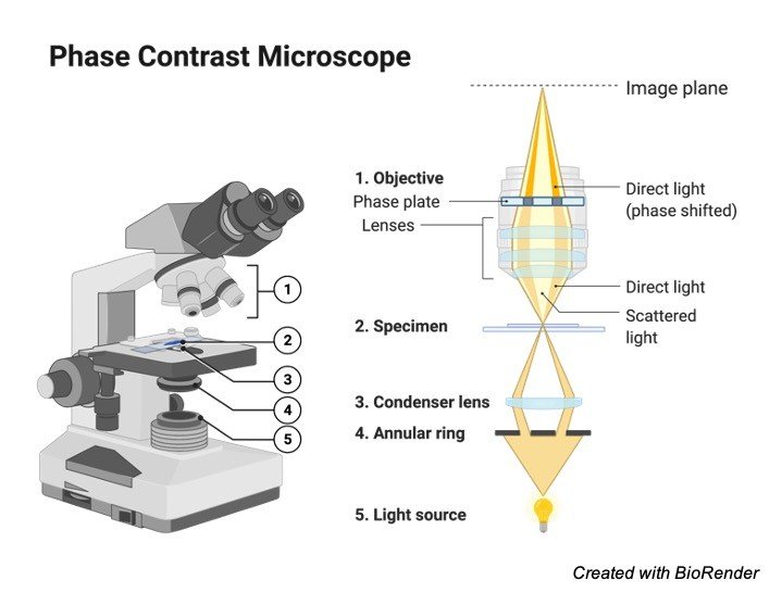
Cytology. Cytology. radiation used to illuminate the specimen ...
Label the microscope — Science Learning Hub All microscopes share features in common. In this interactive, you can label the different parts of a microscope. Use this with the Microscope parts activity to help students identify and label the main parts of a microscope and then describe their functions. Drag and drop the text labels onto the microscope diagram.

General Biology | Carlson Stock Art | General biology ...
STANDARD OPERATING PROCEDURE:

Label the Microscope Diagram | Download Scientific Diagram
9.1: Using Microscopes - Biology LibreTexts

Compound Microscope – Diagram (Parts labelled), Principle and ...
Compound Light Microscope Labeled - ClipArt Best
Compound Microscope Parts, Functions, and Labeled Diagram ...

Compound Microscope Labeled Diagram | Quizlet
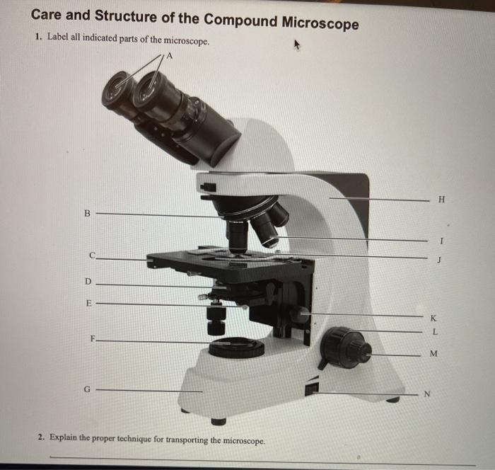
Solved Care and Structure of the Compound Microscope 1 ...

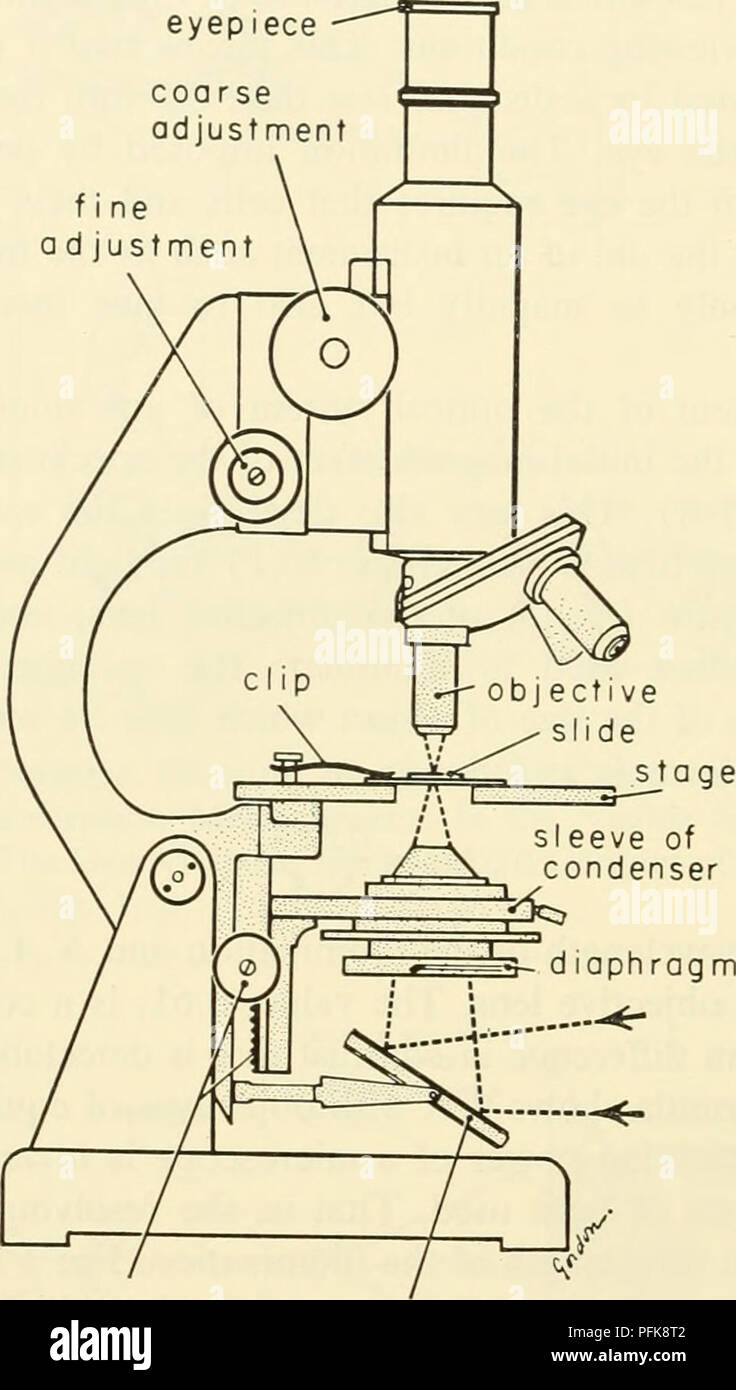
Standard Microscope Diagram | Quizlet
1.2: Microscopes - Biology LibreTexts

Types of Microscopes Archives • Microbe Online

Microscope Parts Review Diagram | Quizlet

Light Microscope- Definition, Principle, Types, Parts ...

Parts of the Microscope with Labeling (also Free Printouts ...

Amscope 40X-2000X Binocular LED Compound Microscope + 5MP Camera + 50 Slides

Microscope study part-2

Compound Microscope Parts, Diagram Definition, Application ...
Compound Microscope Parts, Functions, and Labeled Diagram ...

Compound Microscope- Definition, Labeled Diagram, Principle ...

Simple Microscope - Diagram (Parts labelled), Principle ...
Name Date Sci STANDARD MICROSCOPE DIAGRAM Label only the ...

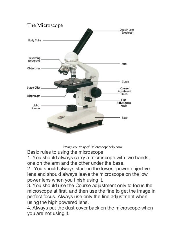
The Microscope
Parts of Microscope, Function, Names & Labeled Diagram ...

This is a common compound microscope. What the labelling D ...
Post a Comment for "39 standard microscope labeled"